
GE Vscan Air Ultrasound: Overview Of Handheld Point‑of‑Care Imaging

Handheld point-of-care ultrasound refers to compact, portable imaging devices that allow real-time sonographic assessment at the bedside or in ambulatory settings. These systems typically combine a small transducer with onboard or paired display software, enabling clinicians to visualize anatomical structures and physiological motion without the footprint of a cart-based unit. Devices in this category may use wireless connectivity to tablets or smartphones, and often emphasize rapid setup, battery operation, and simplified imaging presets intended for focused assessments rather than full diagnostic studies.
Key characteristics of these handheld systems include probe form factor, image processing algorithms, battery life, and the software interface for image display and storage. Some models use conventional piezoelectric transducers while others adopt silicon-based or single-probe architectures that attempt to cover multiple frequency bands. Data security, image export formats, and integration with local workflows can vary across manufacturers, and regulatory clearance for specific clinical uses is typically device- and jurisdiction-dependent.
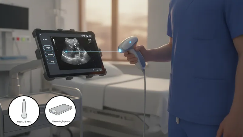
- GE Healthcare Vscan Air — a wireless, pocket-sized transducer that pairs with a mobile display; designed for focused point-of-care imaging and rapid scans.
- Butterfly iQ — a single-probe device that connects to mobile devices and uses semiconductor-based transducer technology intended to cover multiple exam types.
- Philips Lumify series — a software-and-probe approach that pairs phased-array and linear probes with mobile apps for targeted imaging workflows.
When comparing handheld units, image resolution, probe coverage, and software features often guide selection. Image quality can be sufficient for focused assessments such as cardiac window visualization, lung sliding, or limited abdominal surveys, while broader diagnostic studies may still rely on full-size systems. Connectivity options may include Wi‑Fi, Bluetooth, or wired connections to mobile devices. Storage and export support for DICOM or proprietary formats affects how images enter patient records. Regulatory approvals and intended use statements often define which clinical tasks a device may be used for in a given jurisdiction.
Workflow integration typically examines how a handheld ultrasound fits into triage, bedside assessment, and follow-up workflows. Some settings use these devices to augment physical exam findings quickly, potentially reducing time to initial assessment. Integration points include image annotation, automatic measurement tools, and cloud or local archiving. Institutions often consider data governance, user access levels, and compatibility with electronic health record systems when adopting handheld ultrasound into clinical pathways.
Device operation practices include probe handling, contact technique, and selection of imaging presets for different tissue types. Users may employ high-frequency linear presets for superficial structures like vessels and soft tissues, and lower-frequency phased-array or curvilinear presets for deeper abdominal or cardiac views. Battery management, probe disinfection protocols, and routine software updates are practical factors that may influence availability and image consistency across shifts. Training programs typically emphasize hands-on scanning and image interpretation competencies rather than formal device endorsement.
Clinical contexts where handheld ultrasound is often applied include focused cardiac exams, lung assessments for pleural motion, vascular access guidance, and limited abdominal checks for free fluid. These applications rely on focused protocols and may use simplified scoring or measurement approaches. Limitations include smaller display size, potential trade-offs in penetration or resolution, and the need to recognize when a comprehensive diagnostic study on a full-featured system is appropriate. Users should treat handheld imaging as an adjunctive assessment tool within a broader diagnostic workflow.
In summary, compact wireless ultrasound devices are designed to enable targeted, point-of-care sonographic assessment through portable transducers and mobile display solutions. They may streamline focused evaluations and offer connectivity features that support image sharing and archiving, while also presenting considerations for image quality, workflow integration, and governance. The next sections examine practical components and considerations in more detail.