
Laser Eye Surgery: Understanding Procedures, Techniques, And Common Outcomes

Laser-based refractive procedures are a group of ophthalmic techniques that aim to change the way the eye focuses light, typically by reshaping the cornea or altering internal optics. These procedures use controlled laser energy or laser-assisted tools to adjust corneal curvature or tissue profile, with the intent of reducing dependence on corrective lenses. The underlying concept involves precise measurements of refractive error and corneal anatomy, followed by a targeted tissue modification performed under local anaesthesia. Outcomes can vary by technique, eye anatomy, and healing response, so the aim is typically to improve unaided visual acuity rather than to promise a specific result.
Practitioners select among several procedural approaches based on diagnostic measurements, corneal thickness, and patient preferences. Some approaches create a thin corneal flap before applying laser energy, while others work on the corneal surface without a flap. Different laser platforms and guidance systems—such as femtosecond and excimer sources, and wavefront- or topography-guided treatments—may be used to plan and execute corrections. Informed consent and pre-procedure assessment usually cover potential outcomes, risks, and the possibility that additional adjustments may be considered later.
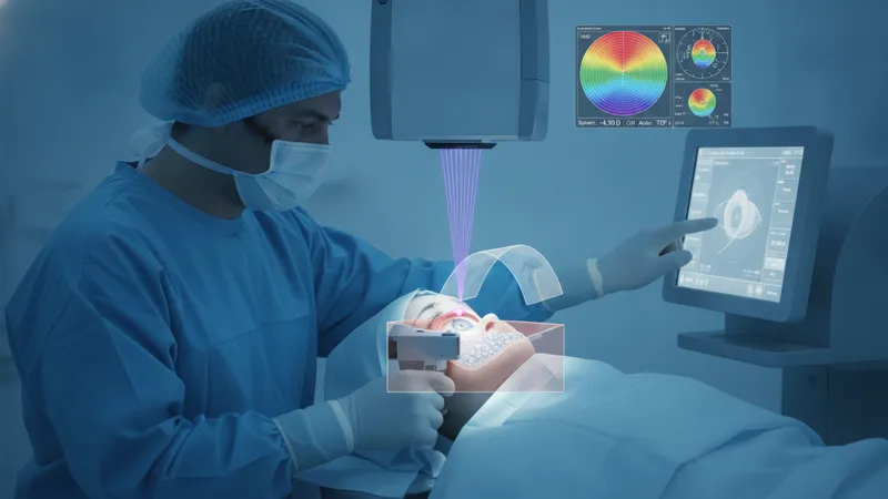
- LASIK: A flap-based procedure where a femtosecond laser may create a corneal flap and an excimer laser reshapes the underlying stroma; typically described for correction of myopia, hyperopia, and astigmatism.
- PRK (Photorefractive Keratectomy): A surface ablation method in which the corneal epithelium is removed and the excimer laser reshapes the cornea; may be used when flap creation is less desirable due to thin corneas or other factors.
- SMILE (Small Incision Lenticule Extraction): A minimally invasive lenticule removal technique performed with a femtosecond laser that creates and extracts a small lenticule through a keyhole incision, commonly applied for myopic corrections with astigmatism in some cases.
Comparisons among these procedures often focus on the structural approach to the cornea, wound healing patterns, and typical recovery timelines. For example, flap-based methods can allow faster visual recovery in many patients, while surface ablation methods may avoid flap-related considerations but often have a longer initial recovery period. Lenticule extraction aims to reduce surface disruption but requires specific laser platforms. When comparing techniques, clinicians typically evaluate corneal thickness, the degree and stability of refractive error, occupational or lifestyle factors, and diagnostic imaging results to identify plausible options rather than to assert a single preferred approach.
Laser and diagnostic technologies influence planning and potential outcomes. Instruments such as corneal topographers, tomographers, and aberrometers measure curvature, elevation, thickness, and higher-order aberrations that may affect visual quality. Treatment planning often integrates these data with manifest and cycloplegic refractions to create a customized ablation profile. Wavefront-guided and topography-guided protocols may aim to address pre-existing optical irregularities, and different laser platforms vary in pulse frequency, spot size, and delivery patterns, which can affect tissue interaction and procedural timelines.
Eligibility considerations commonly include stable refractive error over time, adequate corneal thickness for the intended correction, absence of progressive corneal disease, and reasonable ocular surface health. Age, pregnancy or breastfeeding status, systemic conditions that affect wound healing, and certain autoimmune diseases can be relevant to candidacy. Pre-procedure counseling typically covers these factors, along with the need for a comprehensive eye examination, diagnostic testing, and a discussion of realistic outcomes, recognizing that response to surgery may vary and enhancements may sometimes be considered.
Potential outcomes and risks are routinely part of pre-procedure discussion. Many patients experience reduced dependence on spectacles or contact lenses, but individual results may vary; common transient effects can include fluctuations in vision, ocular dryness, glare, or halos. Less frequent outcomes may include regression toward prior refractive error, infection, or structural weakening in susceptible corneas. Monitoring and post-procedure care aim to detect and manage these issues promptly, with further clinical evaluation guiding any subsequent steps.
In summary, laser-based refractive techniques use targeted corneal or lenticule modifications guided by diagnostic imaging to alter focus and reduce spectacle dependence. The three representative examples above illustrate different interfaces with corneal tissue and distinct recovery profiles; treatment selection typically rests on diagnostic findings, anatomical constraints, and patient circumstances. The following sections examine practical components and considerations in more detail.