
GE Vscan Air Ultrasound: Overview Of Handheld Point‑of‑Care Imaging

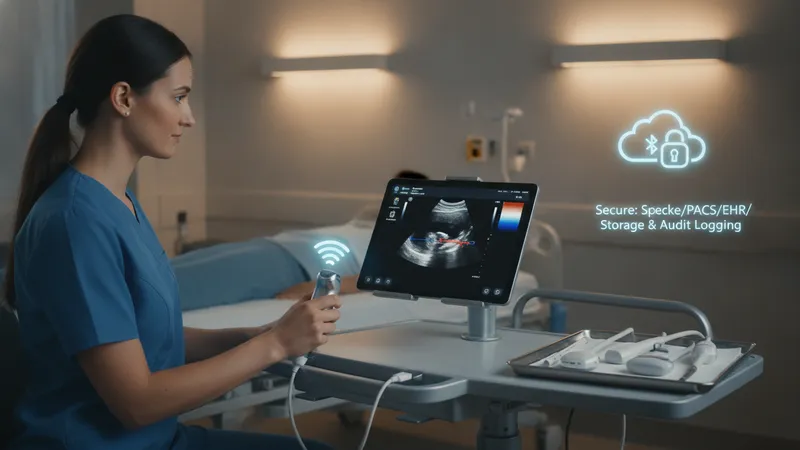
Connectivity and imaging capabilities in handheld ultrasound systems
Connectivity options shape how handheld ultrasound images are displayed, stored, and shared. Devices that pair wirelessly with mobile phones or tablets often rely on Wi‑Fi or Bluetooth protocols; some also support direct wired connections for faster image transfer or charging. Cloud-based storage and vendor-hosted archives may be available, but local on-device storage and export to institutional PACS or EHR systems are common integration considerations. Data encryption, user authentication, and audit logging are technical factors that institutions often evaluate for secure image handling.
Imaging capabilities vary by probe technology and onboard processing. Some handheld probes combine multiple frequency bands into a single transducer, enabling a range of superficial to deeper tissue views, while others use dedicated probes for linear or phased-array imaging. Software features such as harmonic imaging, speckle reduction, and basic measurement tools may improve visual interpretation for focused exams. Color flow or pulse-wave Doppler capabilities are less common in the smallest devices but may be present in higher-tier handheld systems.
Image quality and frame rate often affect the range of clinical tasks that are appropriate for a handheld unit. For dynamic assessments like cardiac motion or lung sliding, temporal resolution can be important. For vascular access or soft tissue work, spatial resolution at superficial depths may be prioritized. Operators typically consider the balance between penetration depth and resolution when selecting presets; device documentation frequently outlines recommended exam types and expected image performance characteristics.
Interoperability with existing clinical IT systems is a practical consideration for deployment. Support for standard formats such as DICOM, HL7 interfaces, or secure file transfer protocols can influence how efficiently images and reports are incorporated into patient records. Device vendors may provide APIs or integration toolkits to assist with automated workflows, but institutions often require validation of data flow, retention policies, and compliance with applicable privacy regulations prior to routine clinical use.